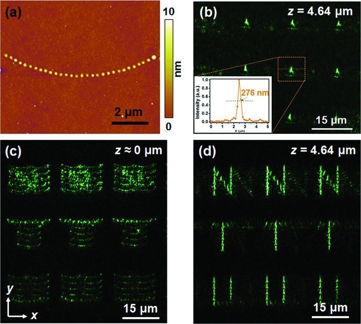
Figure 5.

(a) AFM image of the curved structure with an RC r = 8.6 μm and a 300-nm interspacing. Image (b) is the TIRM image of the convex structures at z = 4.64 μm. The inset depicted in (b) is the transverse intensity profile of focusing spot of the convex structure. (c) and (d) are the TIRM images of the designed structures, which are arranged by the curved structures in (a), observed at z = 0 μm and z = 4.64 μm, respectively. The incident SPP wave propagates in the +y direction.
