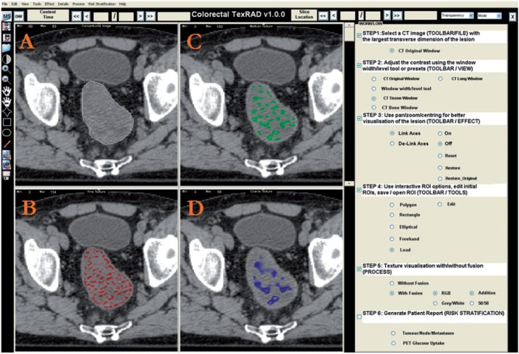
Figure 1.
Conventional CT image of a CRC lesion (A) and corresponding images selectively displaying fine (B), medium (C) and coarse (D) texture obtained by using LoG filter values of 1.0 (width, 4 pixels or 3.9 mm), 1.5 (width, 6 pixels or 5.9 mm) and 2.5 (width, 12 pixels or 11.8 mm), respectively (courtesy of Professor Ashley Groves, Institute of Nuclear Medicine, University College Hospital, London, UK).

