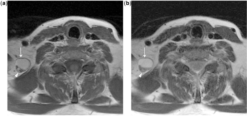
Figure 11.
A 41-year-old woman with treated papillary carcinoma and a cystic nodal recurrence. She was initially treated with thyroidectomy and a central neck dissection followed by ablative 131I therapy. Serum thyroglobulin levels were not increased on follow-up, but a palpable low neck mass was evident. (a) Axial T1-weighted MRI demonstrates a rounded hyperintense lesion (arrow) with a posterior solid nodule (arrowhead) anterior to the right trapezius muscle corresponding to level Vb. The lesion has similar signal intensity to adjacent fat. (b) Axial T2-weighted MRI shows the lesion to be T2 hyperintense (arrow) except for the solid posterior nodule (arrowhead). This was resected and found to be a predominantly cystic papillary thyroid nodal recurrence. The T1 and T2 hyperintense signal likely represents high protein content in the cyst from colloid, thyroglobulin or blood products. Intrinsically hyperintense nodal metastases can be difficult to appreciate on T1 and non-fat-saturated T2 and post-contrast sequences, especially when they are small nodal metastases. Cystic metastases may also be negative on 131I and PET imaging.

