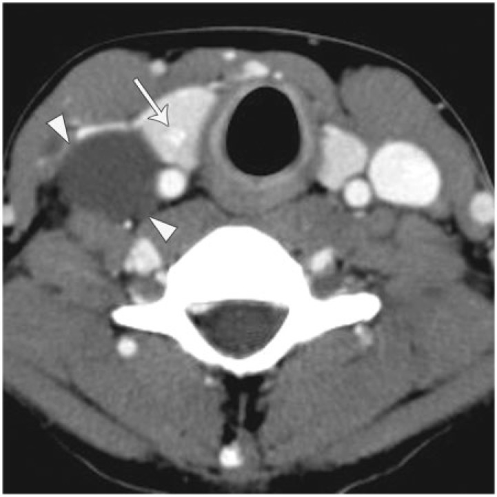
Figure 3.
A 19-year-old woman with papillary thyroid carcinoma presenting with cystic nodal metastases. Axial enhanced CT image shows a radiographically simple cyst (arrowheads) that actually represents a right level IV nodal metastasis. The right internal jugular vein is compressed anterior to the cyst indicating this lesion lies in the carotid space. There is a 1 cm solid primary tumor in the right lobe of the thyroid with fine calcifications (arrow). The differential for a cystic neck mass in a young patient and particularly in a female is a cystic nodal metastasis from thyroid carcinoma, SCCa and a congenital cyst such as a branchial cleft cyst.

