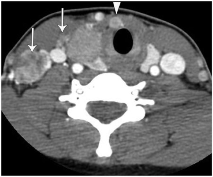
Figure 5.
A 24-year-old woman with metastatic papillary carcinoma including a Delphian nodal metastasis. She presented with a right neck mass. Axial enhanced CT image shows a large mass in the right lobe of the thyroid. There are heterogeneously enhancing right level IV nodal masses (arrows) and an enlarged Delphian node (arrowhead).

