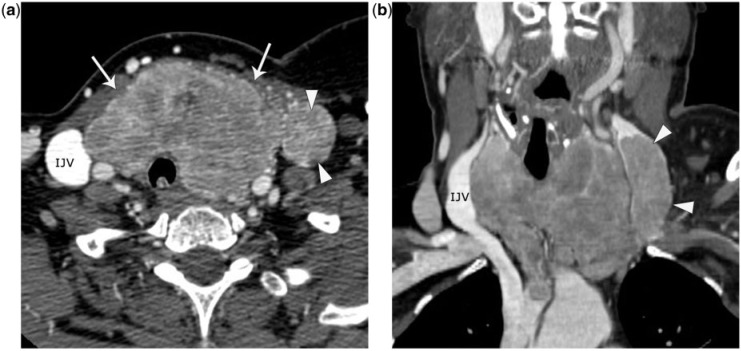
Figure 7.
A 51-year-old woman with follicular carcinoma with venous invasion. She presented with an enlarging neck mass. (a) Axial enhanced CT image demonstrates a heterogeneously enlarged thyroid gland (arrows), displacing the trachea to the right. This was biopsied and determined to be follicular carcinoma. There was no evidence of neck adenopathy, and what resembles a node in the left neck (arrowheads) represents intravenous extension of tumor in the left internal jugular vein (IJV). (b) Coronal reformatted enhanced CT image better delineates extension of tumor in the left IJV (arrowheads).

