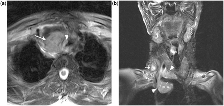
Figure 8.
A 68-year-old woman with papillary thyroid carcinoma with nodal metastatic disease invading the trachea. (a) Axial T2-weighted image shows a T2 hyperintense mass in the right paratracheal region (arrow) with soft tissue signal in the right tracheal cartilage and an intraluminal mass (arrowhead). (b) Coronal T2-weighted image shows the mass encasing the right brachiocephalic artery (BCA) with loss of the fat plane. There is also a right level IV nodal metastasis (curved arrow). She was treated with radioactive iodine and tracheal stenting. Four months later she presented with massive hemoptysis. CT images at presentation showed progression of disease.

