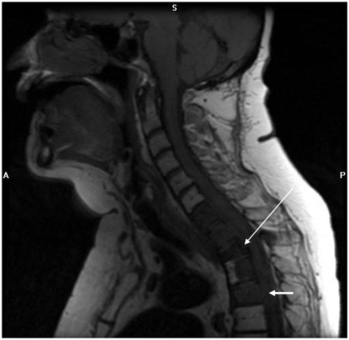
Figure 13.
Sagittal T1-weighted image of the cervical spine in a 61-year-old woman with endometrioid endometrial adenocarcinoma reveals a nearly diffuse abnormal hypointense signal in C7–T4 vertebral bodies with pathologic fracture of T2 (long arrow) and epidural extension posterior to several of the affected vertebral bodies (short arrows).

