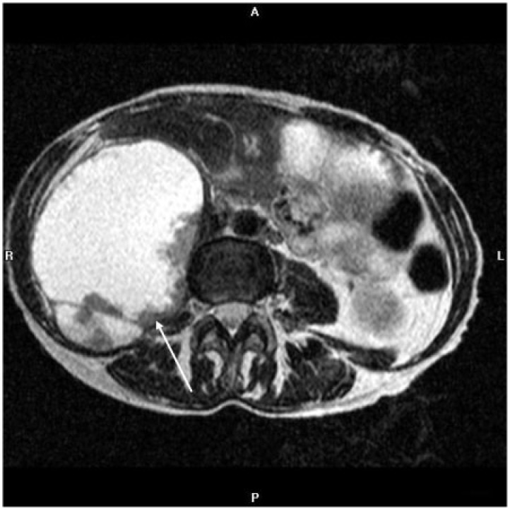
Figure 14.
Axial T2-weighted MR image of the abdomen of a 59-year-old woman with clear cell endometrial adenocarcinoma shows a large, predominately hyperintense mass that expands the right psoas muscle and displaces the bowel anteriorly, with peripheral intermediate intensity components representing tissue (arrow). Resection revealed a fluid-containing endometrial cancer metastasis.

