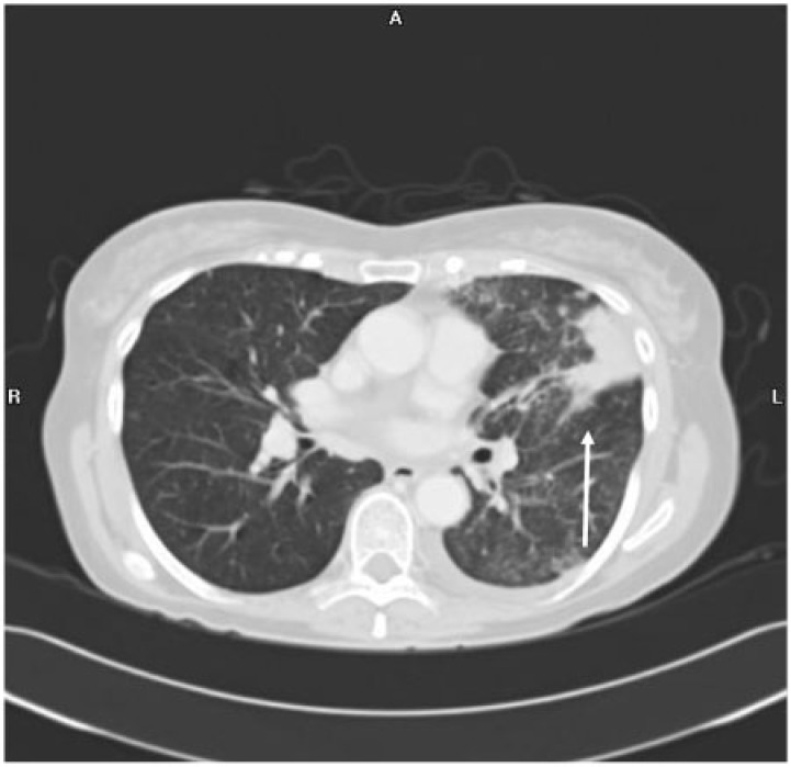
Figure 7.
Axial CT image in a lung algorithm in a 76-year-old woman with clear cell endometrial adenocarcinoma shows a consolidative left upper lobe mass with adjacent nodular septal thickening and peribronchial thickening representing lymphangitic carcinomatosis. Small nodules are seen elsewhere within the imaged lungs.

