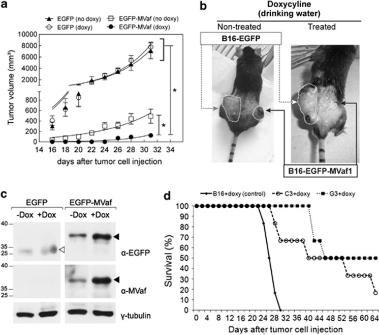
Figure 6.
Effects of MVaf1 expression on tumor growth and animal survival rate in the B16-F10-bearing murine model. (a) Tumor growth rates. Mice bearing EGFP-MVaf1-expressing cells and treated with doxycycline (doxy) did not develop tumors until after the 24-day observation period. Significant differences were verified using an unpaired two-tailed Student's t-test. (*) P<0.05 compared with controls, as indicated on the figure. (b) Representative images of mice bearing tumors of EGFP-MVaf1 and EGFP-transduced B16-F10 cells. The B16-F10 melanoma tumor incidence rate and size were dramatically decreased by EGFP-MVaf (± doxy). Arrows show the site for EGFP-MVaf1-expressing tumor (black) and EGFP-expressing tumor (gray) (n=8/group). (c) Western blots of solid tumor lysates. Lysates were prepared 30 days post inoculation and were probed with commercial anti-GFP antibody (α-EGFP) and laboratory-made anti-MVaf (α-MVaf); γ-tubulin was used as loading control. Black arrowheads indicate EGFP-MVaf1 and white arrowhead indicates EGFP. Analysis shown in a–c were done with whole populations of EGFP-MVaf or EGFP-transduced cells. (d) Survival rates of mice bearing subcutaneous tumors of parental B16-F10 cells or derived clones (C3 and G3) expressing EGFP-MVaf. The survival rates and survival time increased in both C3+doxy and G3+doxy groups (n=6, in observation time point: 66 days post inoculation) compared with the B16+doxy control group (no animal survived beyond 30 days). C3 and G3 are single-cell clones of EGFP-MVaf1 derived from transduced B16-F10 cell

