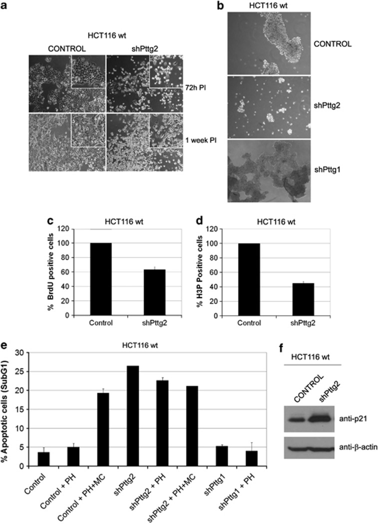
Figure 3.
Silencing of Pttg2 impairs cell adhesion and proliferative capacity of HCT116 cells. (a) Phase-contrast images ( × 10 objective) of Pttg2-silenced HCT116 cells 72 h (upper panels) and 1 week post-infection (PI) (lower panels). Inserts show close-up views of representative cell culture areas. Pttg2-depleted cells show rounder morphology than control cells, indicating a defect in cell adhesion properties. (b) Spheroid formation of Pttg2 and Pttg1-silenced HCT116 (wt) cells in suspension. Seventy-two hours post-infection, cells were transferred to poly-HEMA-coated plates at 6 × 10⁴ cells/ml. After 48 h in suspension, cells were photographed at 10 × . Control and shPttg1-treated cells formed dense spheroids whereas shPttg2-silenced cells adhere to each other very weakly. Representative areas of each culture were selected. (c) Percentage of BrdU-positive cells after shRNA-Pttg2 treatment. HCT116 cells were infected and assayed for BrdU incorporation 72 h post-infection. Bars represent the mean±S.E.M. (d) Analysis of mitotic index in Pttg2-depleted HCT116 cells. Cells were treated with shRNA-Pttg2 lentivirus and incubated with nocodazole 1 μM 72 h post-infection, for 24 h. Percentage of H3P-positive (mitotic) cells was measured by flow cytometry. Bars represent the mean±S.E.M. (e) Percentage of cell death following shRNA Pttg2 or shRNA Pttg1 treatment of HCT116 cells growing under adherent or suspension conditions. Seventy-two hours post-infection, cells were transferred onto control or poly-HEMA-coated (PH) plates in the absence or presence of methylcellulose (MC) and the levels of apoptosis was determined 48 h later by measuring the percentage of cells containing a subG1 DNA content by flow cytometry. Results represent the means of three independent experiments±S.E.M. (f) p21 induction in Pttg2-depleted HCT116 cells. HCT116 cells treated with shRNA-Pttg2 lentivirus for 72 h were harvested and the levels of p21 determined by immunoblotting using specific p21 antibodies. β-Actin was used as loading control

