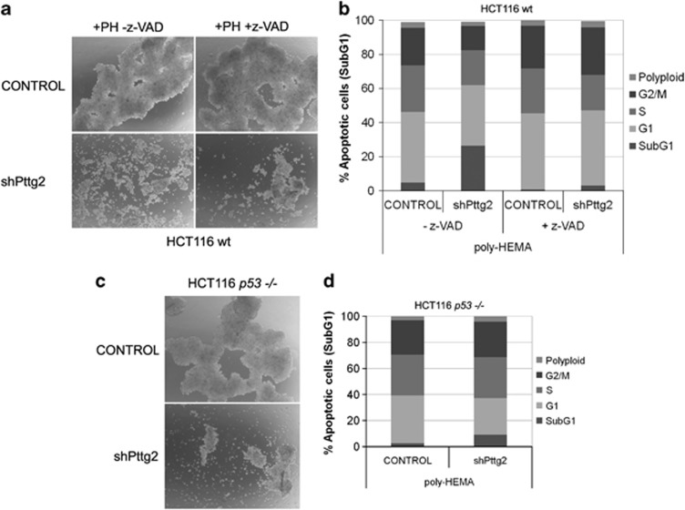
Figure 5.
Loss of cell adhesion in shPttg2-treated cells precedes the onset of apoptosis. (a) Spheroid formation of Pttg2-depleted cells after z-VAD treatment. Wild-type HCT116 cells were infected for 72 h in the presence or absence of the caspases inhibitor z-VAD and the cell–cell adhesion capacity assayed on poly-HEMA (PH)-coated plates. The experiments were repeated three times, and representative areas of each culture are shown. (b) Cell-cycle profile of Pttg2-silenced HCT116 cells growing under poly-HEMA conditions after z-VAD treatment. The percentage of apoptotic cells (subG1) was significantly reduced in the presence of z-VAD, whereas the other cell-cycle phases (G1, S and G2/M) were not modified. Data are representative of two independent experiments. (c) Phase-contrast images to assess multicellular aggregates formation in shPttg2-treated HCT116 p53−/− cells growing on poly-HEMA-coated plates. Images show a representative region of each condition. (d) Analysis of the cell-cycle profile of Pttg2-depleted HCT116 p53−/− cells growing under poly-HEMA conditions. ShPttg2-treated cells lacking p53 showed a reduced percentage of cell death (subG1). The other cell-cycle phases (G1, S and G2/M) remained unaltered. Data are representative of two independent experiments

