Figure 4.
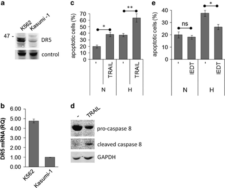
Involvement of the extrinsic pathway in hypoxia-induced apoptosis. (a, b) Routinely cultured Kasumi-1 or K562 (positive control) cells were lysed and proteins subjected to SDS-PAGE and immunoblotting with the anti-DR5 antibody and equalization of protein loading verified on the same membrane (a), or total mRNA was recovered and the relative expression of DR5 calculated by Q-PCR using GAPDH for normalization and Kasumi-1 cells as calibrator (b). (c–e) Kasumi-1 cells were incubated in hypoxia for 4 days in the absence or the presence of 100 ng/ml human recombinant TRAIL (c, d) or 50 μM z-IEDT-FMK caspase 8 inhibitor II (IEDT; e). (c, e) The percentage of apoptotic cells was measured by Annexin V test and flow cytometry. Values represent the average±S.E.M. of data from two independent experiments. The statistical significance of differences was determined by the Student's t-test for paired samples (*P<0.05; **P<0.01; ns: not significant). (d) Cells were lysed and proteins subjected to SDS-PAGE and immunoblotting with anti-caspase 8 antibody. Equalization of protein loading was verified on the same membrane

