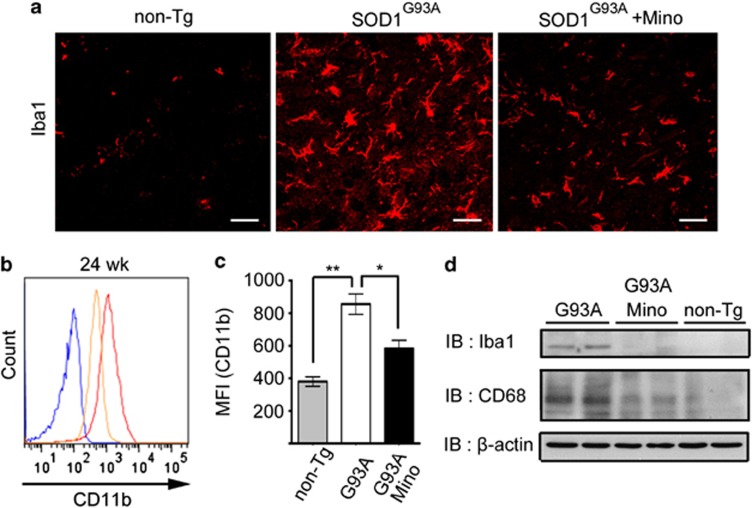
Figure 2.
Minocycline inhibited microglial activation in SOD1G93A mice. (a) The spinal cords of non-Tg, SOD1G93A, and minocycline-treated SOD1G93A mice at 24 weeks (wk) were stained with anti-Iba1 antibody. Bars, 20 μm. (b) A representative profile of CD11b expression at 24 wks. Blue line: non-Tg mice; red line: SOD1G93A mice; orange line: minocycline-treated SOD1G93A mice. (c) Quantitative data on the mean fluorescence intensity (MFI) of CD11b (n=3). Error bars, S.E. **P<0.01, *P<0.05. (d) Lumbar spinal cord lysates from non-Tg, SOD1G93A, and minocycline-treated SOD1G93A mice at 24 wks were subjected to western blotting against Iba1 and CD68 (n=2). β-Actin was used as the internal control

