Figure 6.
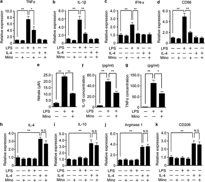
Minocycline selectively inhibited the LPS-induced M1 polarization of microglia. Primary cultured microglia were treated with LPS, IL-4, minocycline, or a combination of these agents. The expressions of M1 (TNF-α (a), IL-1β (b), IFN-γ (c), CD86 (d)) were examined by quantitative RT-PCR. (e) The concentration of nitric oxide induced by LPS in the culture medium was measured after the treatment with minocycline. The concentration of inflammatory cytokines (IL-1β (f) and TNF-α (g)) induced by LPS was measured by ELISA and M2 (IL-4 (h), IL-10 (i), arginase1 (j), and CD206 (k)) markers were examined by quantitative RT-PCR. **P<0.01,*P<0.05. NS, not significant (n=5).

