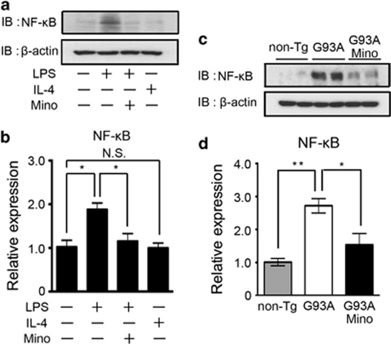
Figure 7.
The LPS-induced upregulation of NF-κB was inhibited by the treatment with minocycline. (a) A representative image of western blotting. Primary cultured microglia were treated by LPS, IL-4, minocycline, or a combination of these agents. Whole-cell lysates were subjected to western blotting. β-Actin was used as the internal loading control. (b) The mRNA expression of NF-κB was analyzed by quantitative RT-PCR in primary cultured microglia. The protein (c) and mRNA (d) expression of NF-κB in the spinal cords of non-Tg, SOD1G93A, and minocycline-treated SOD1G93A mice at 24 weeks were analyzed by western blotting and quantitative RT-PCR, respectively

