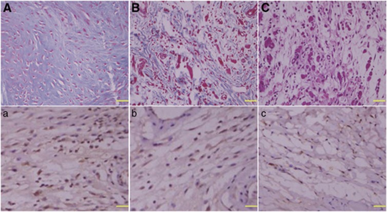
Figure 1.
Representative histological images of collagen tissue and fibroblast proliferation in sciatic nerve anastomosis of rats. The sections obtained from model group (A and a), FK506 group (B and b) and normal control group (C,c) were stained with Masson's trichrome (A, B and C) and anti-TGF-β (a, b and c), respectively. The collagen tissues and fibroblasts appear blue in the sections stained with Masson's trichrome or anti-TGF-β. The density of collagen tissue (B), and the number of fibroblast (b) in the sections of FK506 group were significantly less than those (A, a) of model group. Images × 400 magnified, scale bar 20 μm

