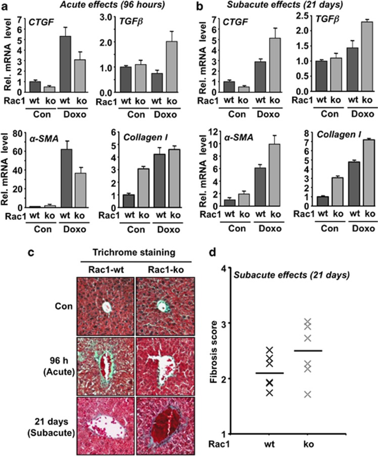
Figure 5.
rac1 influences doxorubicin-induced acute and subacute pro-fibrotic stress responses. (a, b) Rac1 expressing (Rac1-wt) or rac1 deleted (Rac1-ko) animals were treated with a single high dose of doxorubicin (15 mg/kg) (Doxo) and analyzed 96 h later (a) (acute effects) or were repeatedly treated with doxorubicin (3 × 6 mg/kg) and sacrificed 21 days after the first injection (b) (subacute effects). mRNA expression level of selected marker of fibrosis (TGFβ, CTGF, αSMA, collagen I) was analyzed by qRT-PCR. Relative mRNA expression was normalized to the mRNA levels of GAPDH and β-actin and set to 1.0 in non-treated animals. Data shown are the mean±S.D. from triplicate determinations using cDNA generated from pooled RNA samples of n=3–6 mice per group. (c, d) For detection of fibrotic tissue remodeling, trichrome staining (Masson–Goldner) was accomplished as described in Materials and Methods. (c) The photograph shows a representative result. (d) Fibrosis score was analyzed under subacute setting as described in Materials and Methods. Quantitative data shown were obtained from n=6 mice per group. Horizontal bar represents the median

