Figure 4.
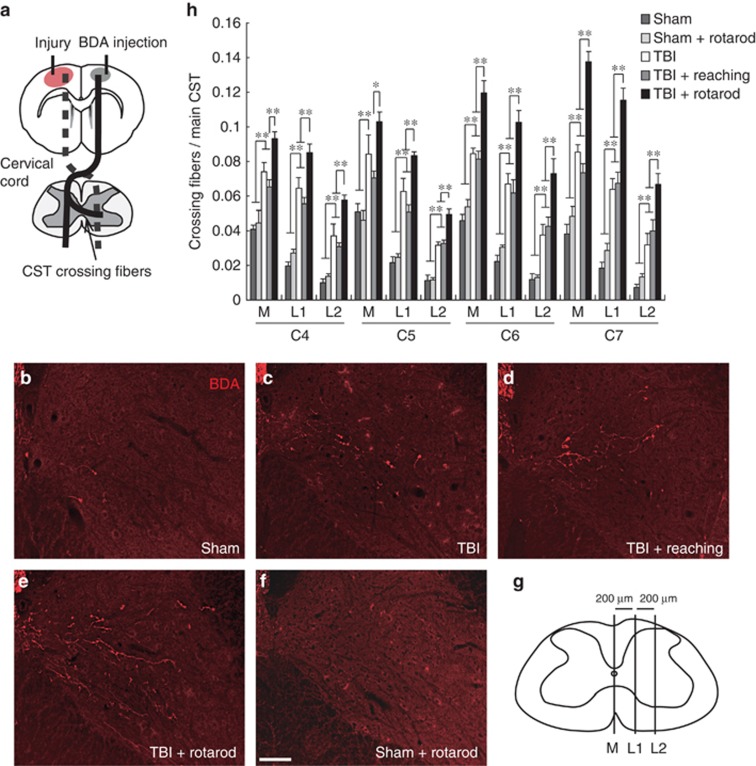
CST rewiring in the cervical cord after rehabilitative training. (a) Diagram of the injury model. The injury to the primary motor cortex leads to the degeneration of corticofugal CST projections (dotted lines). BDA was injected into the contralesional sensorimotor cortex to label intact CST. The arrow shows the rewired crossing fibers from intact CST to the denervated side that are related to functional recovery. (b–f) Representative images of BDA-labeled CST fibers at C7 of the denervated cervical cord in sham (b), TBI (c), TBI+reaching (d), TBI+rotarod (e), and sham+rotarod groups (f) (34 days after TBI). Scale bar: 100 μm. (g) Spinal cord illustration indicating three vertical lines at 200-μm intervals (M, L1, and L2) to create compartments for counting crossing fibers. (h) The number of crossing CST fibers in each compartment (M, M–L1; L1, L1–L2; and L2, lateral to L2) of the denervated side in C4–C7. Two-way ANOVA followed by Tukey–Kramer test, *P<0.05, **P<0.01

