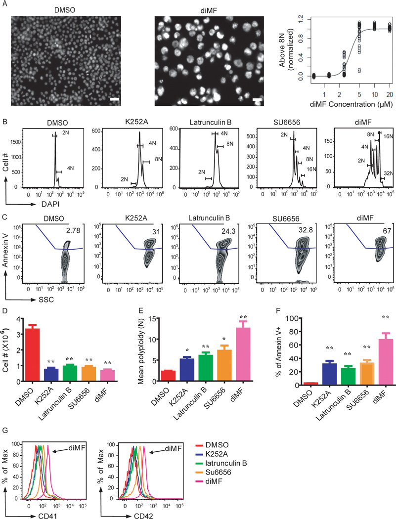
Figure 2. Lead compounds induce polyploidization, expression of differentiation markers, and apoptosis of a human megakaryocytic cell line.
(A) Left, images of Hoechst-stained CMK cells treated with DMSO or diMF. Right, EC50 determination for diMF induction of polyploidization > 8N. Scale bar: 50 µm. (B–G) K252a (5 µM), latrunculin B (5 µM), SU6656 (4 µM) and diMF (5 µM) induced polyploidization (B,E), apoptosis (C,F), proliferative arrest (D) and expression of CD41 and CD42 (G) in CMK cells 72 hr after treatment. Representative flow cytometry plots are shown. Bar graphs depict mean ± SD of two independent experiments conducted in triplicate; * p<0.05, ** p<0.01

