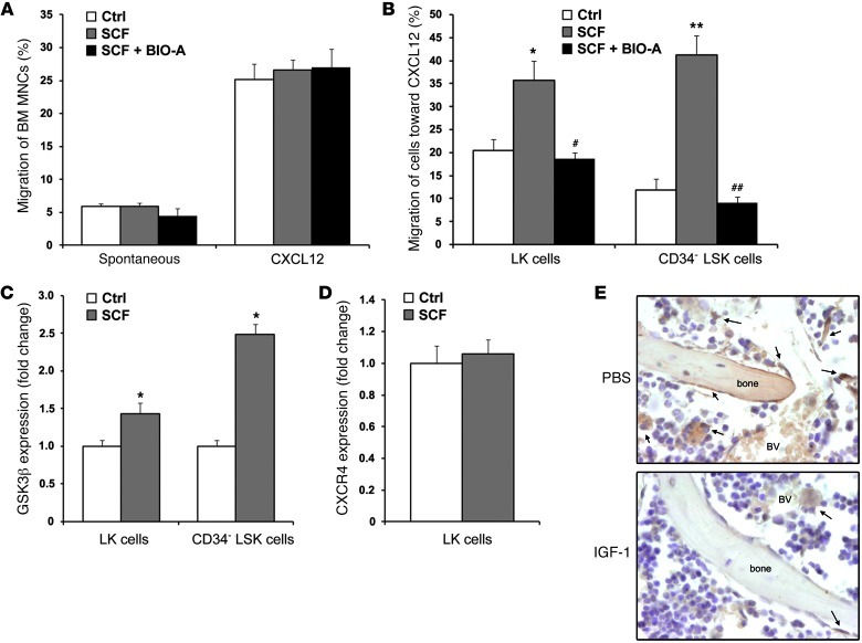
Figure 6. SCF selectively enhances HSPC motility by activating GSK3β in HSPCs.
(A–D) Indicated cells were pretreated or not with 100 ng/ml SCF together with 1 μM BIO-A or equivalent DMSO for 4 hours. (A) BM MNCs were loaded into Transwells. Migration was assessed for 2 hours as being either spontaneous or toward 125 ng/ml CXCL12 (n = 5–6). (B) In addition, LK cells were measured among migrating BM MNCs (n = 3–4), and CD34– LSK cells were measured among migrating Lin– BM cells (n = 8). (C) GSK3β expression (fold change) was determined by flow cytometry in LK cells (n = 5–6), and in CD34– LSK cells (n = 4–5). (D) CXCR4 expression (fold change) was determined by flow cytometry in LK cells (n = 6). (E) Immunohistochemistry for SCF (brown) in the BM after administration of PBS or IGF-1 for 7 consecutive days. Representative images are shown from 3 independent experiments. Original magnification, ×600. Arrows point to SCF-expressing cells. BV, blood vessel or sinusoid. *P < 0.05 compared with control; #P < 0.05 compared with SCF treatment; **P < 0.01 compared with control; and ##P < 0.01 compared with SCF treatment.

