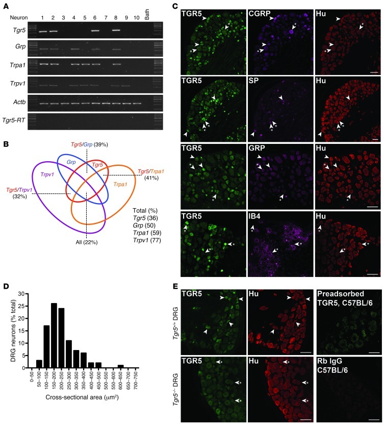
Figure 1. TGR5 expression and localization in mouse DRG.
(A) Single-cell RT-PCR analysis of DRG neurons from C57BL/6 mice. Small-diameter neurons were selected, and Tgr5, Grp, Trpa1, and Trpv1 mRNA was amplified. Results from 10 neurons are shown (78 neurons, 7 mice). Neurons 1, 2, 6, and 8 coexpressed Tgr5, Grp, Trpa1, and Trpv1. No transcripts were amplified from bath fluid. (B) Proportion of small-diameter neurons expressing Tgr5 (36%), Grp (50%), Trpa1 (59%), and Trpv1 (77%). Of the Tgr5-expressing neurons, 39% coexpressed Grp, 41% coexpressed Trpa1, and 32% coexpressed Trpv1. Tgr5, Grp, Trpa1, and Trpv1 were all coexpressed by 22% of small-diameter neurons. (C) Localization of TGR5-IR, Hu-IR, CGRP-IR, SP-IR, GRP-IR, and IB4-FITC binding in DRG (thoracic, lumbar, and sacral) of C57BL/6 mice. Arrowheads denote neurons coexpressing markers; arrowheads with asterisks denote lack of marker coexpression. TGR5-IR was prominently expressed in small-diameter Hu-positive neurons, most of which coexpressed CGRP-IR, SP-IR, or GRP-IR. TGR5-IR was rarely expressed in neurons that bound IB4-FITC. (D) Cross-sectional area of the TGR5-IR population (50-μm2 bins), which indicated that 50% of TGR5-IR neurons were 150–250 μm2. (E) Controls for specific detection of TGR5-IR. TGR5-IR was prominently detected in small-diameter DRG neurons of Tgr5-WT mice (arrowheads). TGR5-IR of small-diameter neurons of Tgr5-KO mice was markedly diminished (arrowheads with asterisks), although the background fluorescence of larger-diameter neurons was retained. Preadsorption of the TGR5 antibody with the receptor fragment used for immunization abolished TGR5-IR in DRG of C57BL/6 mice. There was no staining when the primary antibody was replaced with normal rabbit (Rb) IgG. Scale bars: 50 μm.

