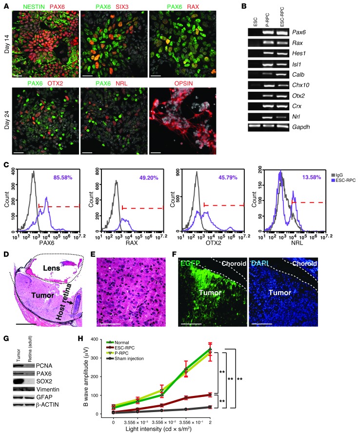
Figure 1. ESC-RPC but not P-RPC transplantation generates ocular tumors.
(A) Immunofluorescence images of costaining of PAX6 with NESTIN, SIX3, and RAX, respectively, in ESC-RPCs at 14 days and costaining of PAX6 with OTX2 and NRL, respectively, or opsin staining alone at 24 days of the differentiation. DAPI is shown in gray. (B) Representative RT-PCR analysis of the expression for retinal markers in ESCs, P-RPCs, and ESC-RPCs. (C) FCM profiles of ESC-RPCs for subpopulations expressing PAX6, RAX, OTX2, and NRL, respectively. (D) H&E staining of the section of an ocular tumor 3 weeks after ESC-RPC injection. (E) The magnified image of tumor tissue in D. (F) A cryosection of a transplanted eyeball containing the tumor tissue from injected ESC-RPCsEGFP (green). (G) Protein levels of several markers in tumor tissues were compared with those in the normal adult retina. (H) The amplitude of ERG b waves at different light intensities is shown for normal mice and SI-treated mice after subretinal injection of P-RPCs, ESC-RPCs, and the cell culture medium (Sham injection), respectively. Mean ± SD; ANOVA; **P < 0.01; n = 10 for each group. Scale bar: 25 μm (A); 500 μm (D); 100 μm (E and F).

