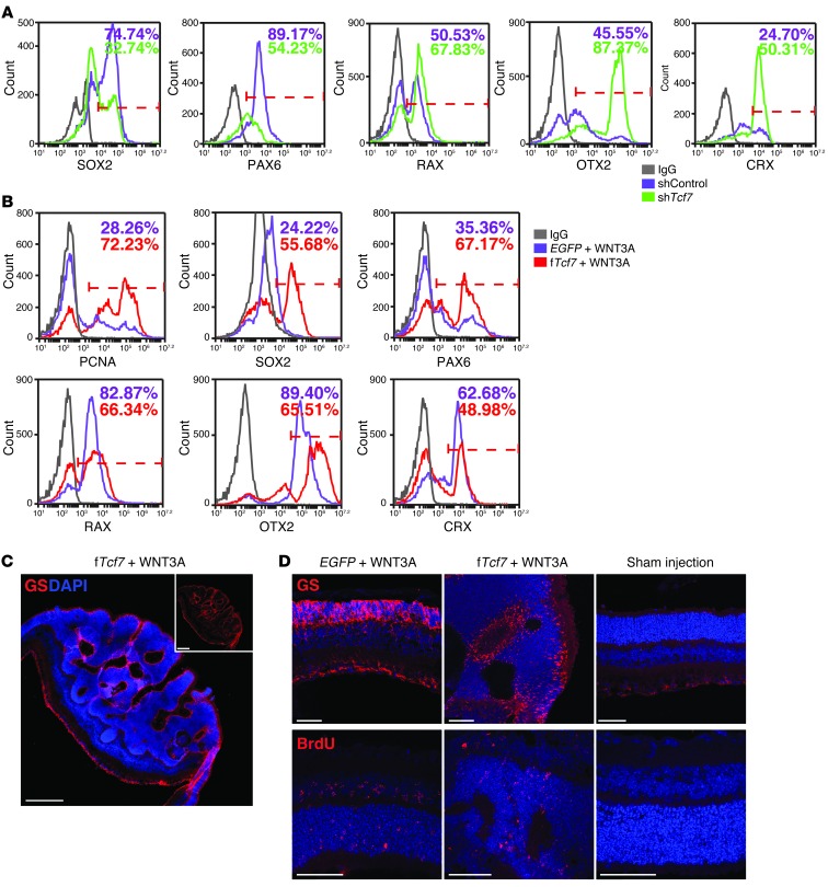
Figure 5. The expression level of TCF7 is directly linked to the state of ESC-RPCs and eye tumor formation in vivo.
(A) FCM analysis to compare the percentage of ESC-RPCs expressing SOX2, PAX6, RAX, OTX2, and CRX, respectively, between shControl and shTcf7-ESC-RPCs. (B) FCM analysis to compare the percentage of P-RPCs expressing PCNA, SOX2, PAX6, RAX, OTX2, and CRX, respectively, between vector-infected and fTcf7-infected P-RPCs in the presence of WNT3A protein. (C and D) Immunofluorescence staining with antibodies against glutamine synthetase (GS) and BrdU in retinae injected with lentiviral fTCF7 and WNT3A protein. Retinae injected with lentiviral EGFP plus recombination WNT3A protein and the medium were used as controls. Scale bar: 50 μm (C); 250 μm (D).

