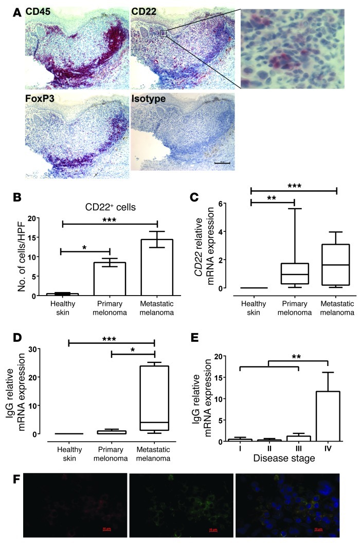
Figure 1. B cells (CD22+) infiltrate melanoma lesions and produce IgG.
(A) Immunohistochemistry showing the presence of lymphocytes (CD45+), mature B cells (CD22+), activated lymphocytes (FoxP3+) (alkaline phosphatase [red], hematoxylin [blue]), and colocalization of all 3 within cutaneous metastases (scale bar: 100 μm; original magnification, ×10). CD22+ cells in melanoma are shown at higher magnification (original magnification, ×40). (B) Significantly increased CD22+ B cell infiltration was measured in primary (n = 6) and metastatic (n = 7) melanoma lesions compared with healthy skin (n = 8). HPF, high-powered microscope field. (C) Comparative real-time PCR showed significantly elevated CD22 expression in primary (n = 10) and metastatic (n = 10) melanomas compared with healthy skin (n = 9). (D) Increased expression of mature IgG mRNA in metastatic melanoma lesions (n = 10) compared with primary melanomas (n = 10) and healthy skin (n = 9) measured by comparative real-time PCR analysis. (E) IgG expression (by comparative real-time RT-PCR) is elevated in melanoma lesions of stage IV patients compared with lesions of stage I–III patients. (F) Immunofluorescent evaluations of IgG+ B cells in human metastatic melanoma lesions (CD22+ B cells in red; left) (IgG+ cells in green; middle) and CD22+IgG+ B cell infiltrates (right). Scale bar: 10 μm; original magnification, ×63. (B and E) *P < 0.01, **P < 0.01, ***P < 0.001, Mann-Whitney U test. (C and D) *P < 0.05, **P < 0.01, ***P < 0.001, Kruskal-Wallis 1-way ANOVA with Dunn’s post-hoc test. Horizontal lines in box plots represent the mean, and whiskers indicate minimum and maximum values

