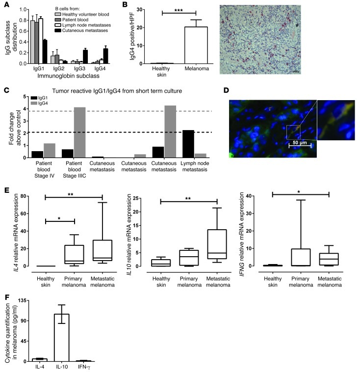
Figure 2. B cells in melanoma lesions are polarized to produce IgG4 antibodies with reactivity against tumor cells.
(A) Polarization of IgG subclasses in B cells derived from metastatic melanoma skin tumor lesions (n = 2), patient lymph nodes (n = 3), peripheral blood from patients with melanoma (n = 6), and from healthy volunteers (n = 4). Cells were cultured ex vivo, and IgG subclass expression profiles were analyzed from culture supernatants by ELISA (mean ± SD; each sample condition tested in triplicate). (B) Immunohistological evaluations confirm significantly increased IgG4+ cell infiltration in melanoma lesions (n = 10) but not healthy skin (n = 10) (***P < 0.001), and representative metastatic melanoma depicts IgG4+ cells (red; hematoxylin [blue]) (scale bar: 20 μm; original magnification, ×10). (C) Reactivity of patient B cell–derived IgG1 and IgG4 antibodies to A375 tumor cells evaluated using a cell-based ELISA. Dashed black and gray lines indicate cut-off points for IgG1 and IgG4 tumor-reactive antibodies, respectively (lines defined as 2 SDs above isotype control antibodies). (D) Immunofluorescent staining of IgG4+ (red) colocalized with S100+ (green) cells in metastatic melanoma (scale bar: 50 μm; original magnification, ×40). (E) Increased expression of IL4, IL10 and IFNG in metastatic melanomas (n = 10) compared with primary melanomas (n = 10) and healthy skin (n = 9) by comparative real-time PCR. Horizontal lines in box plots represent mean, and whiskers indicate minimum and maximum values. *P < 0.05, **P < 0.01, Kruskal-Wallis 1-way ANOVA with Dunn’s post-hoc test. (F) Cytokines IL-4, IL-10 and IFN-γ are secreted in metastatic melanoma lesion ex vivo cultures.

