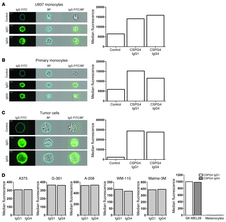
Figure 4. Biological properties of engineered IgG1 and IgG4 antibodies recognizing the melanoma-associated antigen CSPG4.
Anti-CSPG4 IgG1 and anti-CSPG4 IgG4 antibodies bind to receptors on the cell surface of (A) U937 monocytic cells, (B) primary monocytes, and (C) tumor cells. ImageStream and flow cytometric evaluations confirm IgG1 and IgG4 antibody binding to Fcγ receptors (top and middle panels) and also to the antigen CSPG4 (bottom panel) expressed on the surface of SK-MEL-28 melanoma cells. ImageStream images are shown on the left. IgG FITC, cell surface (area indicated by circle on top left of each panel) binding of antibodies on cells; BF, bright-field image of cells; IgG FITC/BF, composite image of antibody and bright-field images, confirming anti-CSPG4 antibody binding to the cell surface. Quantitative analyses are based on the acquisition of 20,000 cells. Control samples for U937 and monocytes were incubated with goat anti-human IgG (Fab′)2-FITC antibody. Control samples for antibody binding to tumor cells were human IgG1 and IgG4 isotype antibodies of irrelevant specificity. (D) Flow cytometric analysis of CSPG4 IgG1 or CSPG4 IgG4 antibody binding to a panel of tumor cell lines or primary human melanocytes. Antibody binding was detected with an anti-human IgG (Fab′)2-FITC, and specific binding was depicted by MFI values above isotype control antibody binding.

