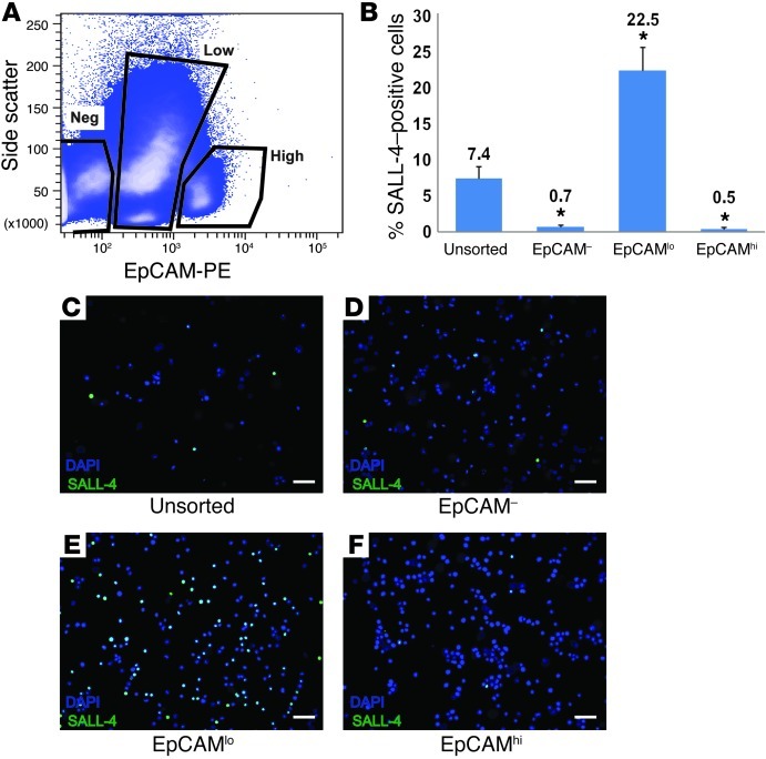
Figure 2. SALL-4–positive spermatogonia are recovered in the EpCAMlo fraction of human testis cells.
(A) Human testicular cells were stained with EpCAM-PE, and 3 populations were identified based upon EpCAM-PE staining intensity and side scatter of incident light. Negative gates were defined by analysis of human testis cells stained using PE-conjugated isotype control antibodies. (B–F) Following sorting, each fraction of cells was fixed and immunocytochemistry assessing SALL-4 expression was performed. Following SALL-4 staining, cells were counterstained with DAPI. Cells from at least 10 independent images were then counted based on DAPI staining and SALL-4 staining, respectively, to determine the percentage of cells expressing SALL-4. An unsorted fraction of cells was also stained with an isotype antibody to control for nonspecific binding to demonstrate specificity. (B) Relative SALL-4 expression in unsorted and EpCAM-sorted fractions. Bars indicate the mean percentage of SALL-4–positive cells (SALL-4–positive cells/total cells) in each fraction. Error bars represent SEM from 3 replicate sorting experiments. *P < 0.001, compared with unsorted cells. (C–F) Representative images from SALL-4 immunocytochemistry of unsorted and sorted fractions. Scale bar: 50 μm.

