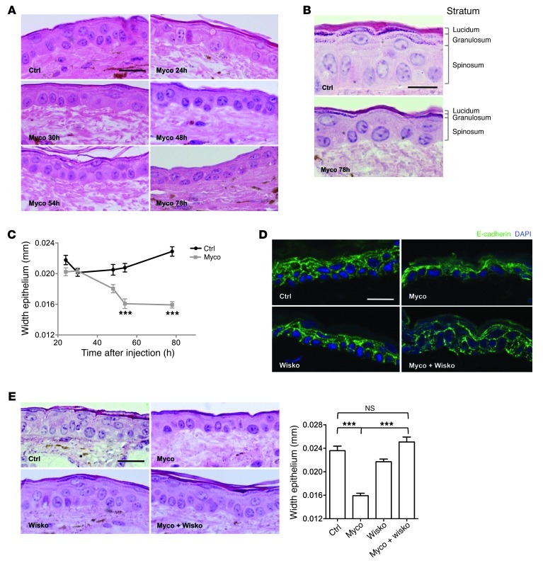
Figure 7. Remodeling of the skin epidermis after mycolactone administration.
(A) H&E-stained sections of ear skin 24, 30, 48, 54, and 78 hours after intradermal injection of vehicle control or 7 nmol (5 μg) mycolactone. (B) Higher-magnification images of control and 78-hour mycolactone sections, showing thinning of all epidermal layers and loss of stratum granulosum. (C) Evolution of epidermal width at the site of injection of mycolactone or vehicle control. Data are mean ± SEM from 10 measurements per mouse in 3 mice. ***P < 0.001, Kruskal Wallis with Dunn post-test. (D and E) E-cadherin staining (D) and H&E staining and mean width (E) of the ear epidermis 78 hours after intradermal injection of 7 nmol mycolactone, 14 nmol wiskostatin, or both compared with vehicle control injection. Shown are representative images from 3 mice per group. Data are mean ± SEM from 10 measurements per mouse in 3 mice. ***P < 0.001, Kruskal Wallis with Dunn post-test. Scale bars: 20 μm (A, D, and E); 10 μm (B).

