
FIGURE 3.
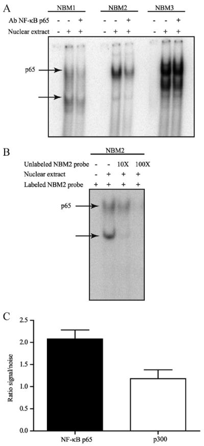
NF-κB p65 binds to the P2Y2R promoter sequence. A, Nuclear extracts from Caco-2 cells were incubated with the putative [γ-32P]ATP-labeled NF-κB p65 DNA-binding site probes NBM1, NBM2, or NBM3 and anti-NF-κB p65 Abs for electrophoretic mobility and supershift assays. DNA-protein complexes were separated from the free probe on a native polyacrylamide gel. Arrow, NF-κB p65 DNAbinding and supershifted complexes. B, Nuclear extracts from Caco-2 cells were incubated with the putative [γ-32P]ATP-labeled NF-κB p65 DNA-binding site probe NBM2 and 10× or 100× unlabeled NBM2 for electrophoretic mobility and competition assays. DNA-protein complexes were separated from the free probe on a native polyacrylamide gel. Results are representative of three independent experiments. NF-κB p65 DNA-binding complex is indicated by the arrow. C, Chromatin was immunoprecipitated with or without rabbit IgG Ab or anti- NF-κB p65 Ab. The re-ChIP assay was performed with anti-p300 Ab following the first immunoprecipitation with anti-p65 Ab. Samples were verified by quantitative RT-PCR analysis with oligonucleotides amplifying the −221 bp to −155 bp region of the P2Y2R promoter and expressed as fold increase over normal rabbit IgG normalized to input. Results are representative of three independent experiments.