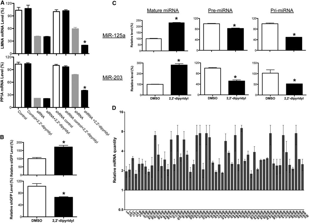
Figure 3. Iron Chelator Promotes the Processing of shRNAs and miRNA Precursors.
(A) Metal chelator enhances only shRNA-mediated but not synthetic siRNA duplex-induced knockdown. Knockdown of each mRNA was graphed as the percentage of mock-treated samples in the presence or absence of metal chelators (2,2′-dipyridyl). Values are mean ± SD for triplicate samples. *p < 0.001. Control was transfected with control siRNA duplex.
(B) Quantitative RT-PCR was used to measure the levels of processed siRNA and shRNA in RNAi-293-EGFP/RFP reporter cells. 5S was used as an internal control. Values are mean ± SD for triplicate samples. *p < 0.001.
(C) Quantitative RT-PCR was used to measure the levels of pri-, pre-, and mature forms of miR-125a and miR-203 in mock- and iron chelator-treated HEK293 cells expressing the primary transcript of miR-125a. The relative expression levels as determined by DDCt analyses are shown. Values are mean ± SD for triplicate samples. *p < 0.001.
(D) Relative quantity of miRNA with consistent changes in expression shown for iron chelator-treated HEK293 cells (transfected HEK293 cells stably expressing pri-miR-125a were used), calibrated to mock-treated cells (mock-treated relative quantity = 1; the mean relative quantity from three pairs is plotted with error represented as a 95% confidence interval).
See also Figures S3 and S4 and Table S1.

