. 2013 Apr 2;2:e00278. doi: 10.7554/eLife.00278

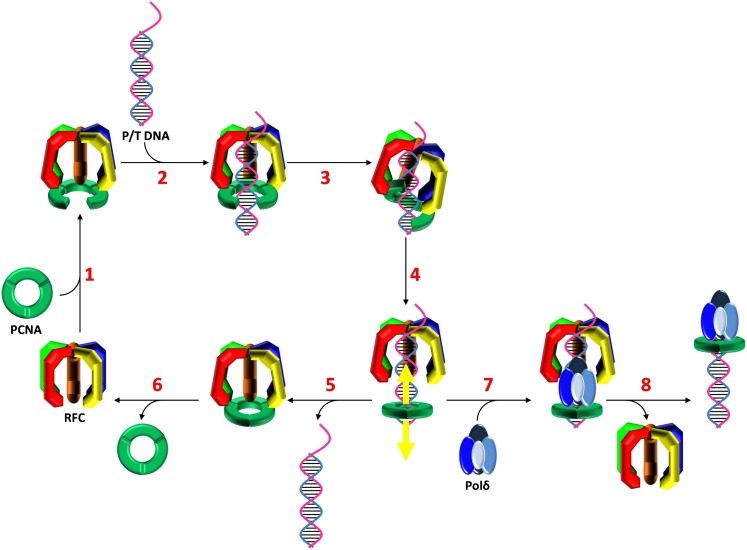
Figure 5. Stepwise assembly of the human DNA polymerase holoenzyme.

(1) RFC•ATP binds PCNA and opens it for assembly onto DNA. (2) The open PCNA•RFC•ATP complex binds to a P/T junction and (3) adopts the notched screw cap arrangement. (4) RFC hydrolyzes ATP, closing the PCNA ring and releasing it onto DNA. (5) In the absence of polymerase, loaded PCNA is unable to ‘escape' from DNA-bound RFC and is unloaded back into solution by RFC. (6) RFC subsequently releases PCNA, exchanges ADP for ATP, and the cycle repeats. (7) In the presence of polymerase, loaded PCNA is ‘captured' from DNA-bound RFC by an incoming polymerase, blocking the unloading activity of DNA-bound RFC by physical occlusion. (8) RFC subsequently dissociates, leaving behind the functional holoenzyme consisting of polymerase and PCNA.

DOI: http://dx.doi.org/10.7554/eLife.00278.022

Figure 5.

Figure 5—figure supplement 1. The human notched screw cap complex.

Figure 5—figure supplement 1.

The DNA footprint of the human RFC•ATPγS•PCNA complex (Tsurimoto and Stillman, 1991) overlaid on the Cy3-P/T DNA substrate. Shown in magenta is the region protected by human RFC. Shown in cyan is the region protected by human PCNA.
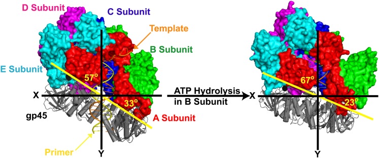
Figure 5—figure supplement 2. The clamp loader gp44/62 of T4 bacteriophage retracts towards the P/T junction upon hydrolysis of ATP and closure of the gp45 clamp ring around DNA.

Figure 5—figure supplement 2.

The open clamp:clamp loader:DNA complex (PDB code 3U60) and the closed clamp:clamp loader:DNA complex (PDB code 3U61) are shown in the same orientation (via alignment of the AAA+ module of the C subunit). Images were generated using Pymol. P/T DNA and gp45 (gray) are shown in cartoon form. The surface of gp44/62 (multicolored) is shown. The clamp loader subunits (A–E), gp45 as well as the primer (orange) and template (yellow) strands of the duplex DNA are indicated.