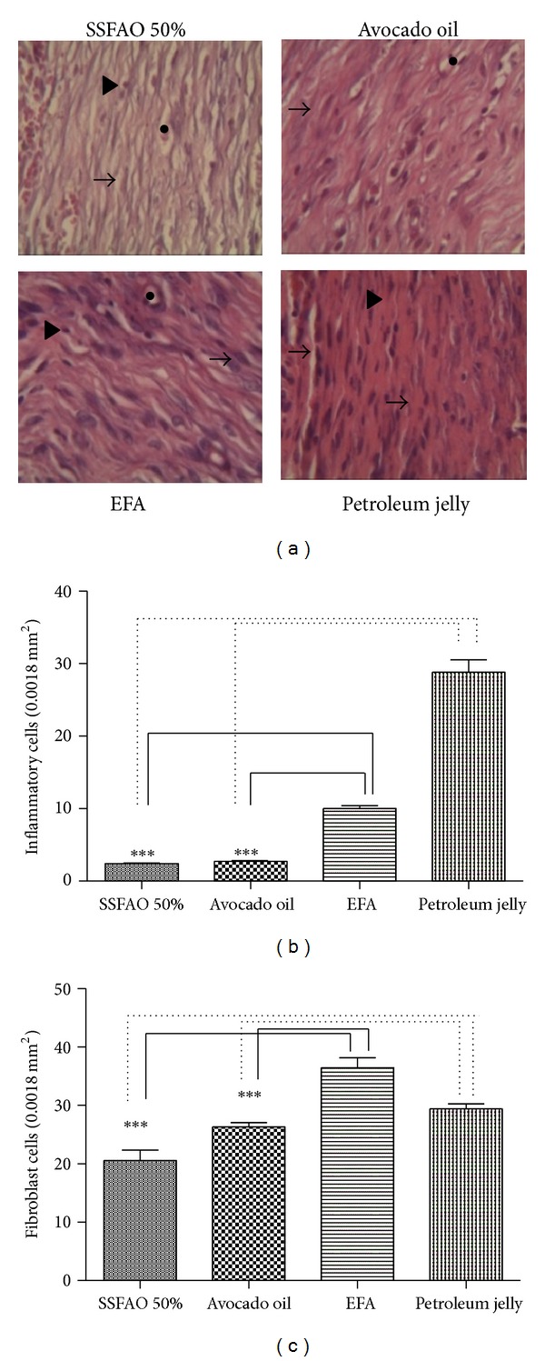
Figure 3.

Histopathologic observation of treated excisinal wound with SSFAO 50%, in natura avocado oil, EFA (control positive), or petroleum jelly (control negative) at the 14th day after operation. Tissue sections were stained with hematoxylin and eosin (400x magnification). (a) Representative images show granulation tissue presenting fibroblasts (arrow) and inflammatory cells (arrowheads) and surrounding capillaries (asterisk). Fewer inflammatory (b) and fibroblast (c) cells are seen in treated excisinal wound with SSFAO 50% or in natura avocado oil when compared to controls. At least 30 different random fields were measured per treatment (n = 6). Data are shown as average ± SEM. *P < 0.05 versus controls; **P < 0.01.
