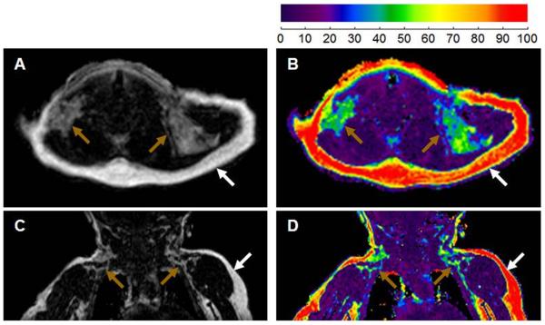
Figure 4.
Imaging characteristics of brown and white adipose tissue in infancy. Axial (A, B) and coronal (C, D) MR views of a 4 month infant depicting both white fat (white arrow) and brown fat (brown arrow) at the level of thoracic inlet. Compared to white fat, brown fat is hypointense/darker in the fat images (A, C), and has a lower fat fraction (green versus red) in the co-registered fat and water images (B, D). (Adapted from Sharp LZ, Shinoda K, Ohno H, Scheel DW, Tomoda E, Ruiz L, Hu H, Wang L, Pavlova Z, Gilsanz V, Kajimura S. Human BAT possesses molecular signatures that resemble beige/brite cells. PLoS One. 2012;7(11):e49452.)

