Figure 2.
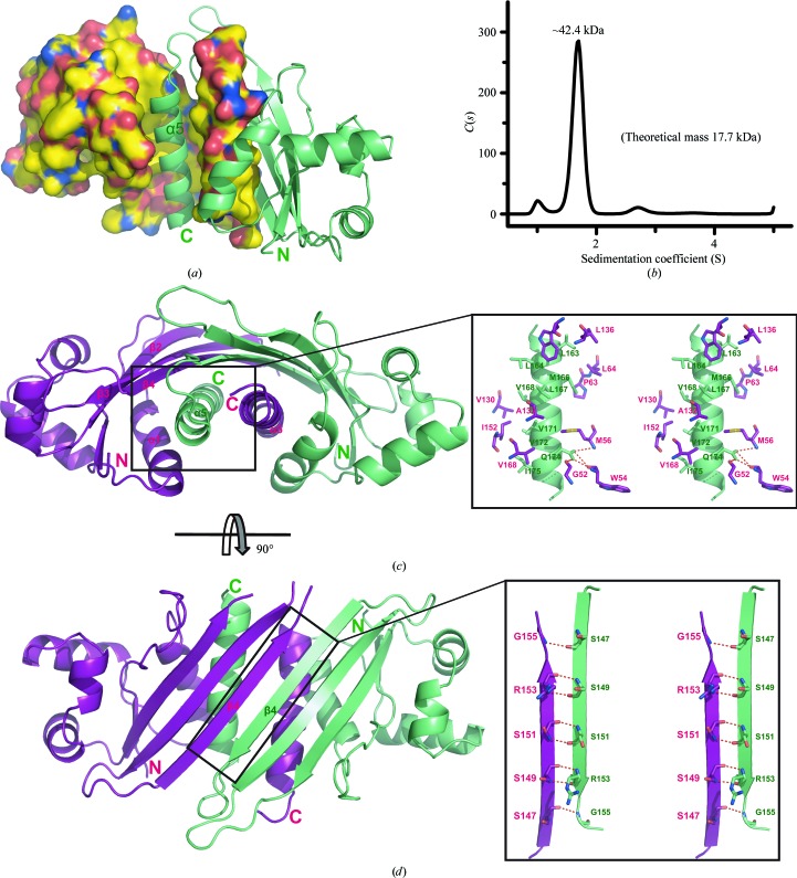
Dimeric packing of GNA1162. (a) The structure of the GNA1162 dimer. Molecule A is shown as a surface representation. C, N, O and S atoms are coloured yellow, blue, red and orange, respectively. Molecule B is shown as a ribbon representation and is coloured green. (b) SV analysis of the GNA1162 protein (residues 26–180). The difference between the theoretical molecular mass (35.4 kDa) and the SV fit may arise from the molecular shape and sample homogeneity. (c) Interface of α5 (green) and the hydrophobic groove (magenta) formed by α1, α5 and β2–β4. (d) Interface of the β4 strands of both molecules. Molecule A is coloured magenta and molecule B is coloured green. Hydrogen bonds are shown as dotted red lines.

