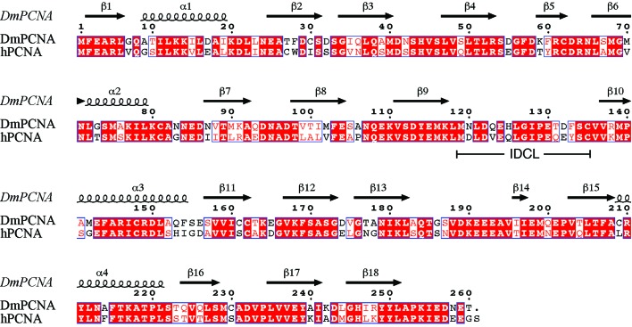
Figure 2.
Sequence alignment of DmPCNA and hPCNA was performed with ClustalW2 (Goujon et al., 2010 ▶; Larkin et al., 2007 ▶) and ESPript (Gouet et al., 1999 ▶). The secondary structure is shown according to the DmPCNA structure. Residues that are indentical in the two species are highlighted with a red background and highly conserved residues are coloured red; all of these residues are boxed.

