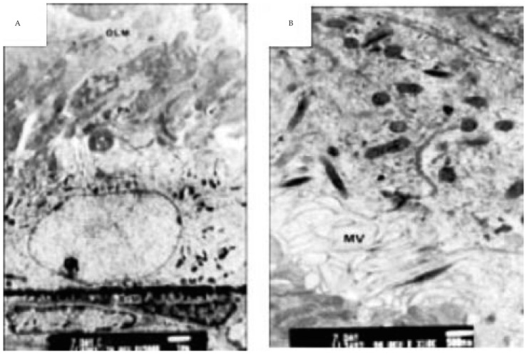
Figure 3. Transmission electron micrographs of control retina of 7 days old offspring rats.
A: showing retinal pigmented epithelial cells with underlying basement membrane. The cytoplasm is rich in mitochondria, ribosomes and smooth endoplasmic reticulum. The apical part is characterized by radial arranged microvilli adjacent to macrophagosome and newly formed inner segment of photoreceptors (×75 00). B: showing branched microvilli (MV) of pigmented epithelium, phagosome and newly formed inner segment of photoreceptors (×13 000).

