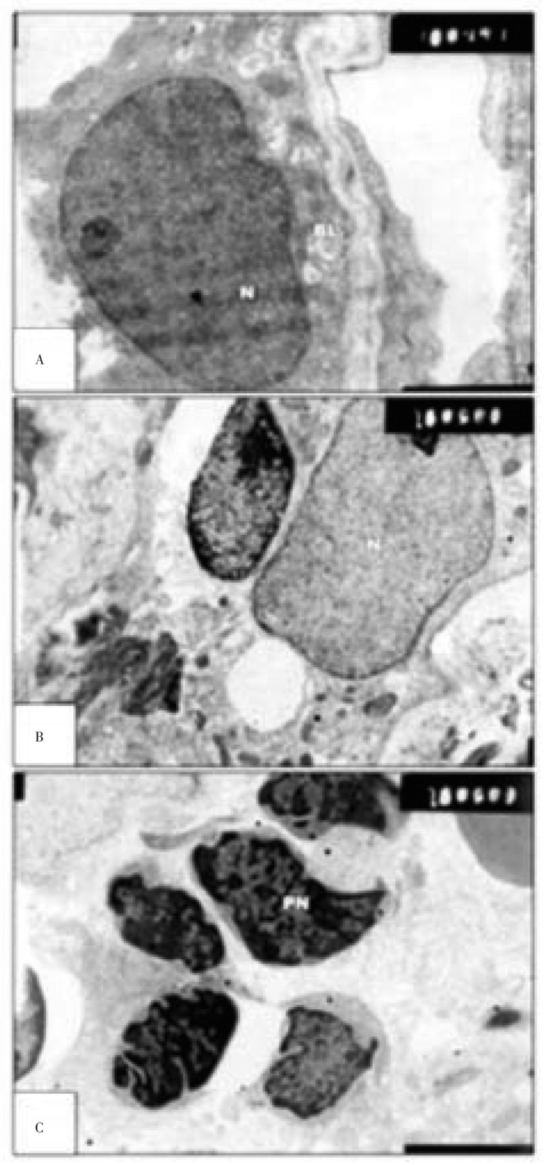
Figure 4. Transmission electron micrographs of experimental retina of 7 day old rats.
A&B: showing malformed pigment cell. Their cytoplasm enclosed by numerous vacuoles (V) and phagosomes. Inner segment appears degenerated in many of them. C: showing massive degeneration of nuclear cells building nuclear layer. The nuclear cells appeared with convoluted and pyknotic nuclei (PN) and electron-dense chromatin material (A&B×13 000; C×7 500).

