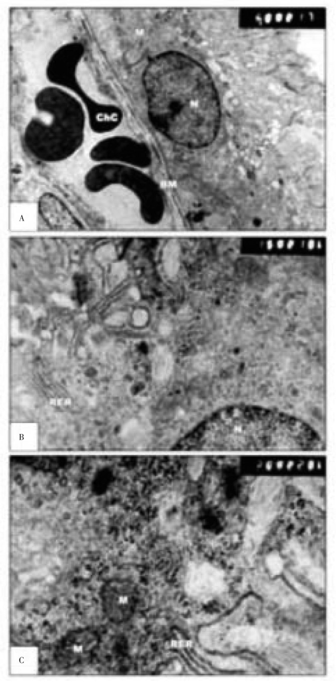
Figure 6. Transmission electron micrographs of experimental retina of 14 days old rats.
A&B: showing pigmented epithelium with vacuolated cytoplasm, vesiculated rough endoplasmic reticulum and fragmented rough endoplasmic reticulum. The choriocapillaris appear swollen. C: showing degenerated outer segment of photoreceptors (A&B×13 000; C×20 000).

