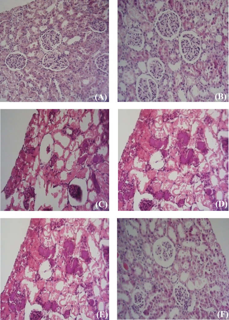
Fig. 3.
Renal histological microphotographs. Male (A, C and E) and female (B, D and F) kidney sections were taken from sham-operated rats groups (A and B) or ischemic-reperfusion groups (C and D) or ischemic-preconditioned groups (E and F). Sham-operated; control group are normal kidney tissues, normal histological characteristic of glomeruli and tubules and corpuscle’s space were present in male (A) and female (B). In male ischemic-preconditioned group (E), there was moderate tubular dilatation with loss of nuclei in most of tubule, without swelling, luminal congestion (e.g., moderate diffuse interstitial edema and moderate dilatation the tubular structure), focal glomerular necrosis and extent free space in corpuscle and predominates over morphological features of apoptosis (e.g., severe chromatin condensation and cell shrinkage). In female Ischemic-preconditioned group (F), there was mild tubular dilatation with loss of nuclei in some of tubule, with swelling, luminal congestion (e.g. mild diffuse interstitial edema and mild dilatation the tubular structure), focal glomerular fragmentation and extent free space in corpuscle and predominates over morphological features of apoptosis (e.g., moderate chromatin condensation and mild cell shrinkage). In male ischemic-reperfusion group (C), there was severe tubular dilatation, focal glomerular necrosis, and extent free space in corpuscle, loss of nuclei, chromatin condensation and cell shrinkage. In female ischemic-reperfusion group (D), there was severe tubular dilatation, moderate focal glomerular necrosis, and moderate extent free space in corpuscle, moderate loss of nuclei, mild chromatin condensation and mild cell shrinkage.

