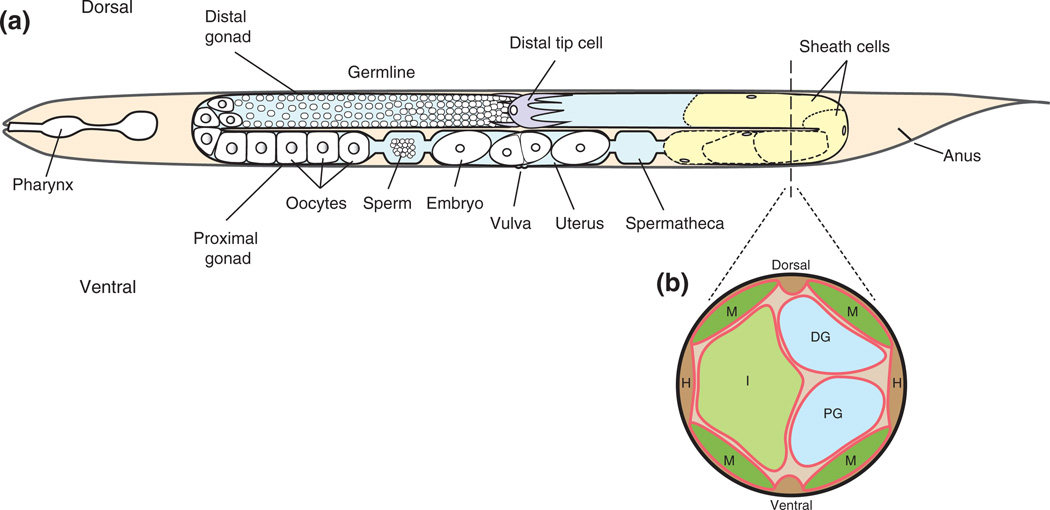
FIGURE 1.
Schematic representation of the adult hermaphrodite. Gonadal structures are featured in a lateral view4 (a) and a cross-sectional view (b). (a) The anterior gonad arm (left) depicts germ cells in the distal gonad; proximal gonad structures are labeled. The posterior arm (right) depicts the somatic structures of the gonad including a distal tip cell (purple) and sheath cells (yellow). The dotted line running through the posterior gonad arm represents the approximate location of the schematic cross-sectional slice. (b) The cross-sectional view depicts the basement membranes (red) that surround the body wall muscles (M), hypodermis (H), intestine (I), distal gonad (DG), and proximal gonad (PG). This schematic is based on an electron micrograph.

