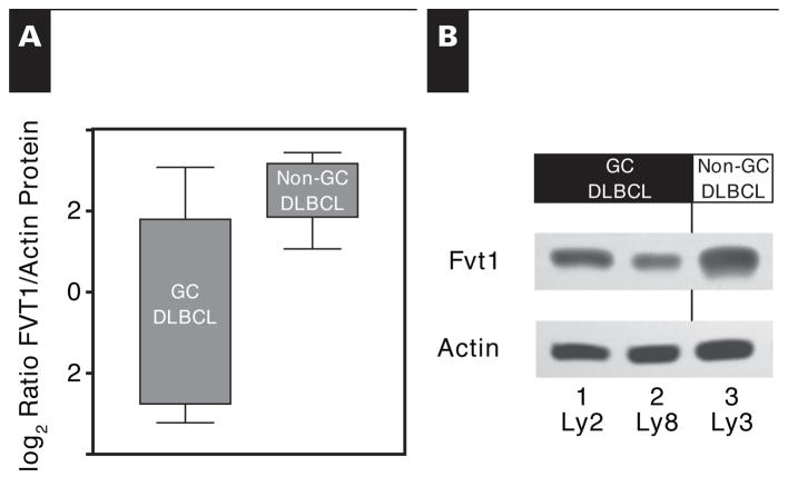
Figure 2.
Amount of follicular lymphoma variant translocation 1 (FVT1) protein as detected by Western blot. A, Amounts of FVT1 protein and actin in 12 selected patient samples were detected by Western blot and quantified by densitometry. Although not statistically significant (P = .12), the trend for FVT1 protein parallels the gene expression findings, with decreased amounts in the germinal center–type cases relative to those of non–germinal center–type cases. B, Amounts of FVT1 protein and actin were detected by Western blot for 3 representative diffuse large B-cell lymphoma cell lines. Those of the germinal center type (Ly2 and Ly8) contain a decreased amount of FVT1 relative to one of the non–germinal center type (Ly3). Boxes represent the second and third quartiles, while whiskers represent the full range of values.

