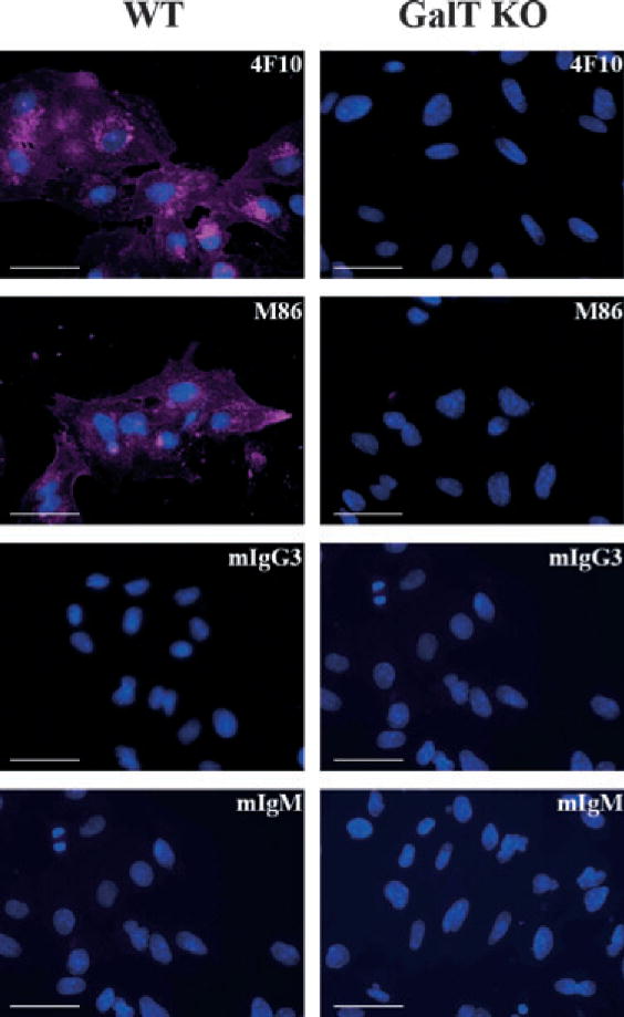
Fig. 2.

Uniform distribution of αGal in wild-type pig aortic endothelial cells. PAEC WT (left column) and GalT KO (right column) were grown directly in 96-well plates; fixed/permeabilized before staining with the 4F10 and M86 mAb or matching isotype controls: IgM and IgG3, respectively, as indicated in the top-right corner of the figures; and analyzed by Olympus fluorescent IX71 microscope fluorescent microscope. Overlay pictures of Evans blue channel in violet and DAPI channel in blue. Bar corresponds to 50 μm. Representative images of four different staining with the different pig cell lines. GalT KO, α1,3galactosyltransferase knockout; mAb, monoclonal antibodies; PAEC, pig aortic endothelial cell; WT, wild-type.
