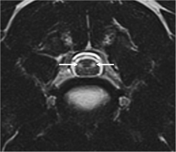
Figure 1.

Transverse T2-weighted MR images of the cervical spinal cord at the level of the C4-C5 intervertebral disc space. The images show well-demarcated, ovoid, hyperintense signals with a bilateral, symmetrical appearance in the region of the lateral funiculi (arrow).
