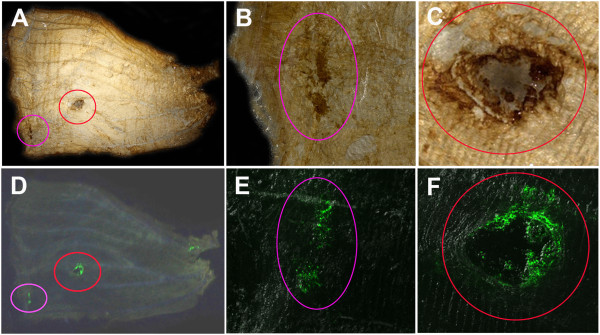
Figure 6.

Immunohistochemical detection of GFP in developing J. orithya pupal wings with anti-GFP antibody. A fourth-day pupal wing infected with 2.0 × 10⁴ pfu/mL baculovirus vector (2.0 μL, 18-24 hours post-pupation) and treated with anti-gp64 antibody (2.0 μL, 18-24 hours post-infection) is shown. Immunohistochemical DAB staining and GFP fluorescent signals overlapped with each other. (A-C) Immunohistochemical DAB staining using anti-GFP antibody. Two major regions indicated by circles were stained in A, and they are magnified in B and C. (D-F) GFP fluorescence signals from the same wing in A-C. Two major regions indicated by circles showed fluorescence, and they are magnified in E and F.
