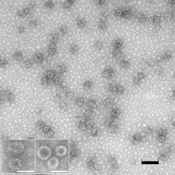
Figure 3.
Electron microscopic view of VP1-survivin VLPs assembled in vitro. The VLP preparation was transferred to carbon-coated Formar copper grids (300 mesh) and stained with 5% uranyl-acetate. The preparation contained multiple irregularly shaped VLPs as well as pentameric structures (black bar=100 nm). The inset depicts (left) VP1-survivin VLPs at higher magnification, and (right) regularly shaped VP1 VLPs expressed in and purified from the yeast S. cerevisiae (bars=50 nm).

