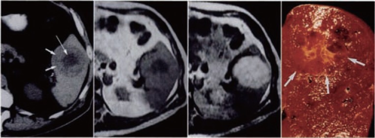
Figure 1.
Splenic haemangioma. Unenhanced CT scan shows a round splenic lesion with lower attenuation in the centre (arrow) than in the periphery. Weighted MR image shows that centre has low signal intensity but periphery is nearly isointense in the spleen. Lesion becomes bright on T2 weighted MR image. Cut surface of gross specimen shows an unencapsulated hemorrhagic lesion (arrows) with central stellate scar. Source: D.Disler, F.Chew, Radiologic-Pathologic Conferences of the Massachusetts General Hospital.

