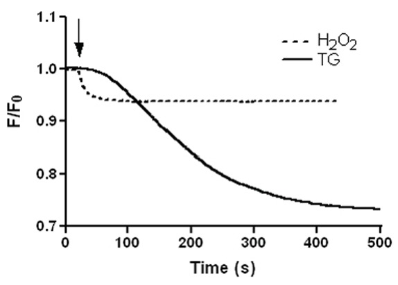
Figure 5.

H2O2 causes a loss of mitochondrial staining by calcein. Calcein-loaded cells in the presence of 1 mM Cl2Co were stimulated with 100 μM H2O2 or 1 µM thapsigargin (TG). Changes in calcein fluorescence were detemined as shown under “Material and Methods” and are expressed as fractional changes of emitted fluorescence (F/F0). Traces are representative of 5–6 independent experiments.
