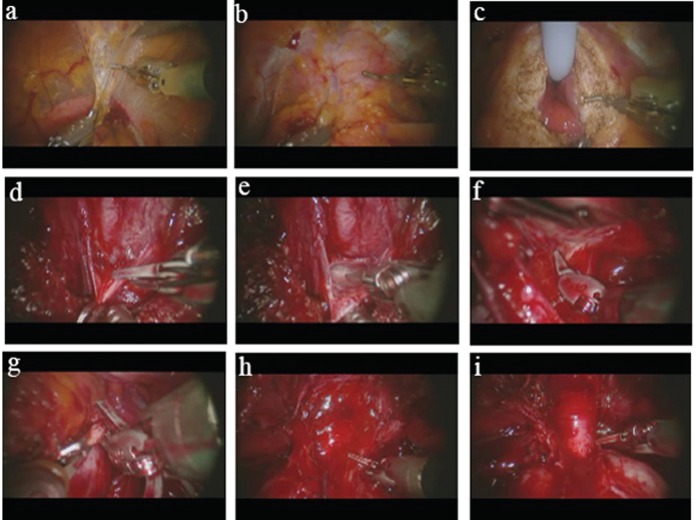
Figure 1.
Photographs of selected steps of our robotic prostatectomy technique. (a) Dissection of the space of Retzius using the Maryland bipolar forceps in the left hand and the cautery hook in the right. The bladder can be seen in the bottom portion of the picture beneath the bipolar forceps; (b) The prostate has been cleared of its fatty covering and the anterior bladder neck (being held by the bipolar forceps) is about to be opened with the cautery hook; (c) After the anterior bladder neck has been opened, the Foley catheter is pulled anteriorly with the fourth arm (ProGrasp forceps, not shown in photo), giving exposure to the lateral and posterior portions of the bladder neck; (d) With the prostate lifted anteriorly with the fourth arm, the Denonvillier's fascia is grasped with the bipolar forceps and cut with the cold scissors (bladder neck seen in foreground); (e) The pearlywhite tissue that is characteristic of the plane of tissue between the layers of Denonvillier's fascia is shown here; (f) The neurovascular bundle is gently dissected off the poserolateral surface of the prostate, shown here on the left side (prostate is being held up by left hand bipolar forceps); (g) The endopelvic fascia is opened towards the apex along the contour of the prostate; (h) The dorsal venous complex is divided with the pneumoperitoneum raised to 20 mm Hg; (i) The apex of the prostate is shown here with the urethra skeletonized and about to be divided close the prostate to preserve maximum urethral length.

